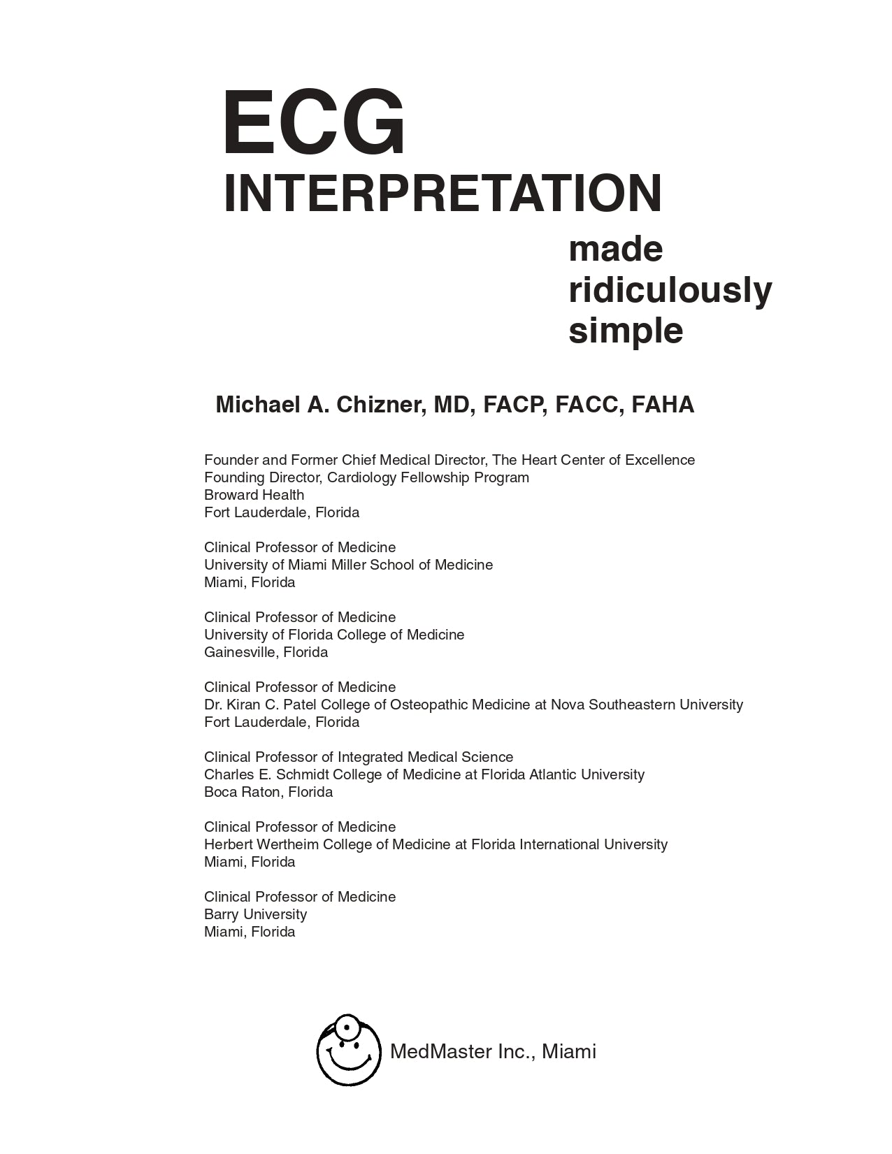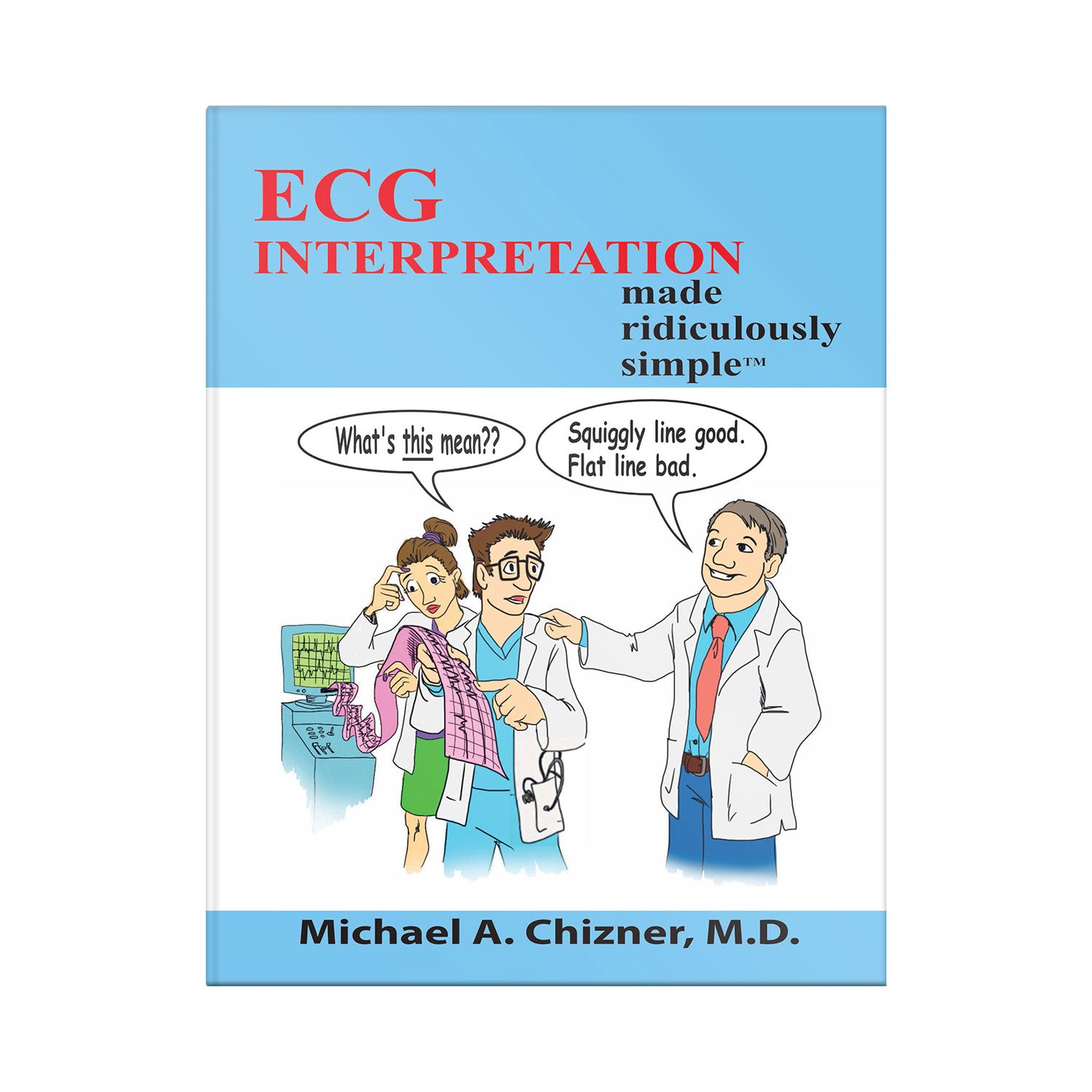
ECG Interpretation Made Ridiculously Simple
Also available on
Description
Author(s)
Michael A. Chizner, M.D.
Michael A. Chizner, M.D., renowned cardiologist, Founder and former Chief Medical Director of The Heart Center of Excellence of Broward Health, was recently named in the Top 1% in the Nation in the first Top Doctors List compiled by U.S. News & World Report. Dr. Chizner graduated with highest honors from Cornell University Medical College. He received his medical residency training at the New York Hospital-Cornell Medical Center, and his cardiology training at Georgetown University, where he received the Distinguished Alumnus Award. He is Board Certified in Internal Medicine and Cardiology, and is a Fellow of the American College of Physicians, American College of Cardiology, and American Heart Association. He is a Clinical Professor of Medicine at the University of Florida College of Medicine, the University of Miami Miller School of Medicine, Nova Southeastern University College of Osteopathic Medicine, the Charles E. Schmidt College of Medicine at Florida Atlantic University, and Barry University. Dr. Chizner has served as Chairman and Vice Chairman of the Florida Board of Medicine. He has also served on the Editorial Advisory Boards of national cardiology journals, and has been Director and Keynote Speaker at national and regional medical education conferences. He has written and edited numerous articles and books in cardiology. His widely read textbook, Clinical Cardiology Made Ridiculously Simple is currently being used in medical schools throughout the U.S. and abroad. Dr. Chizner spearheaded the establishment of the Heart Center of Excellence at Broward General Medical Center, overseeing an exceptional roster of experienced cardiologists and cardiovascular surgeons, whose clinical expertise, superior outcomes, and strongly positive patient satisfaction ratings earned Broward General Medical Center the distinction as the only high performing hospital in Broward County for cardiology and heart surgery, according to U.S. News & World Report.
Details
Pages: 142
Publication: Edition 1 (June 15, 2020)
Language: English
ISBN: 9781935660422 eISBN: 9781935660620
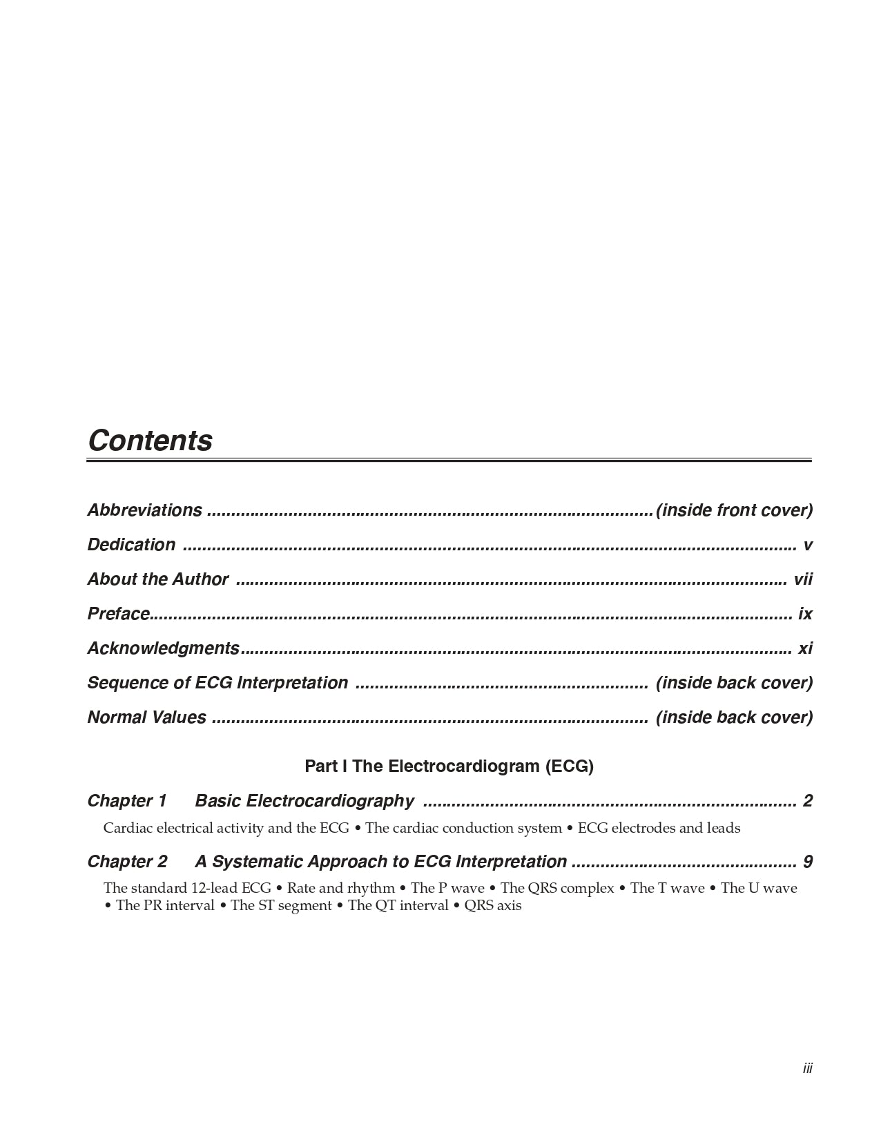
Table of contents
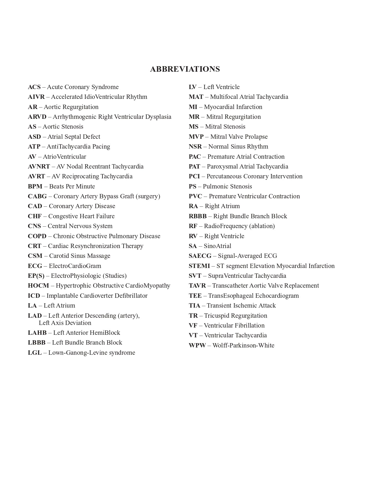
Abbreviations
Dedication
About the Author
Preface
Acknowledgments
Sequence of ECG Interpretation (inside back cover)
Normal Values (inside back cover)
Part I The Electrocardiogram (ECG)
Chapter 1 Basic Electrocardiography
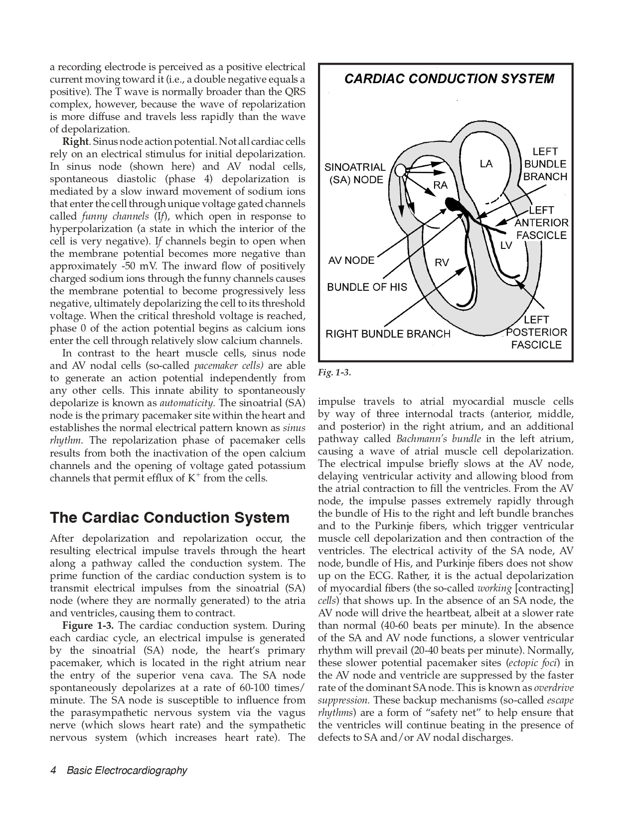
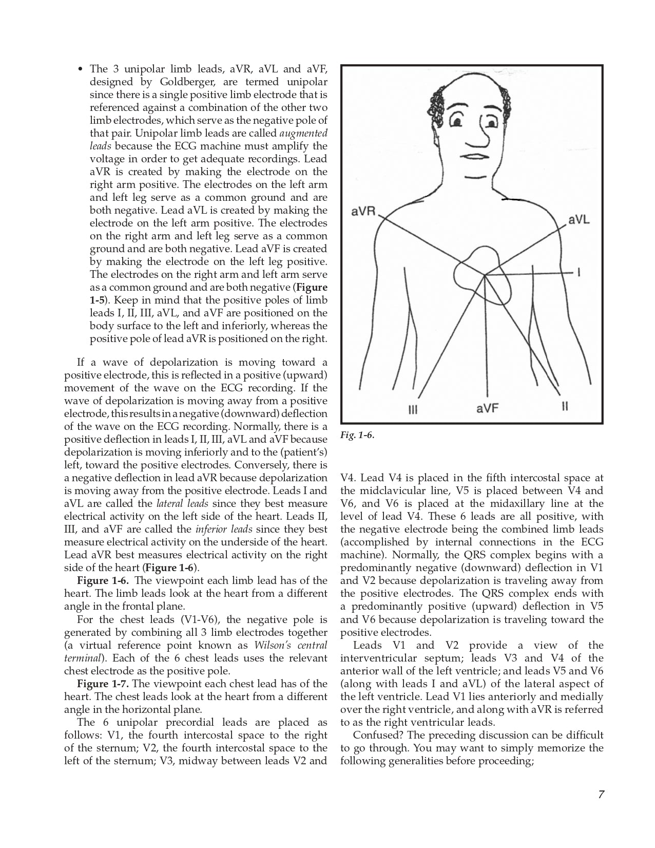
Cardiac electrical activity and the ECG • The cardiac conduction system • ECG electrodes and leads
Chapter 2 A Systematic Approach to ECG Interpretation
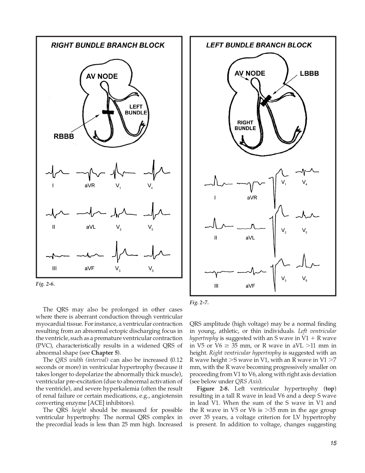
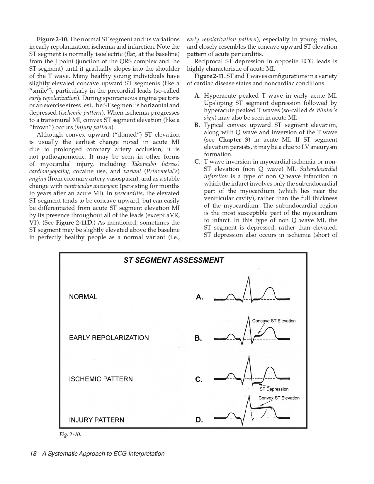
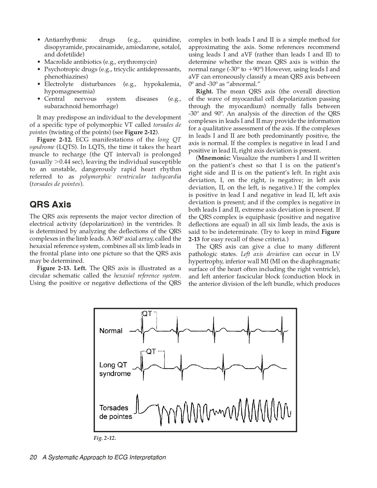
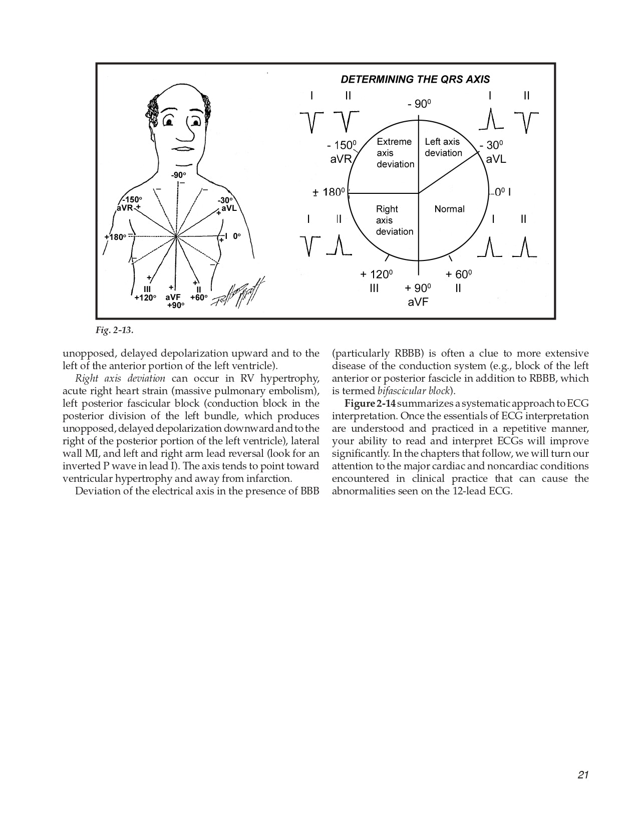
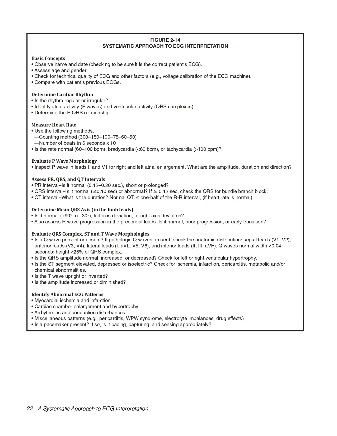
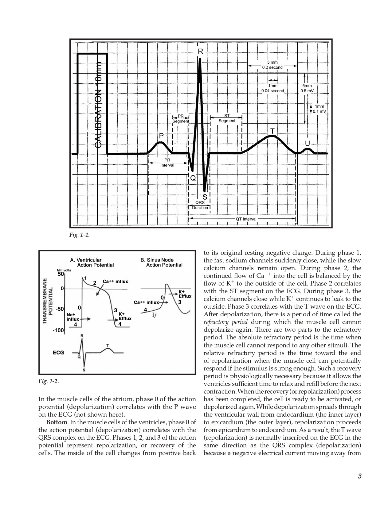
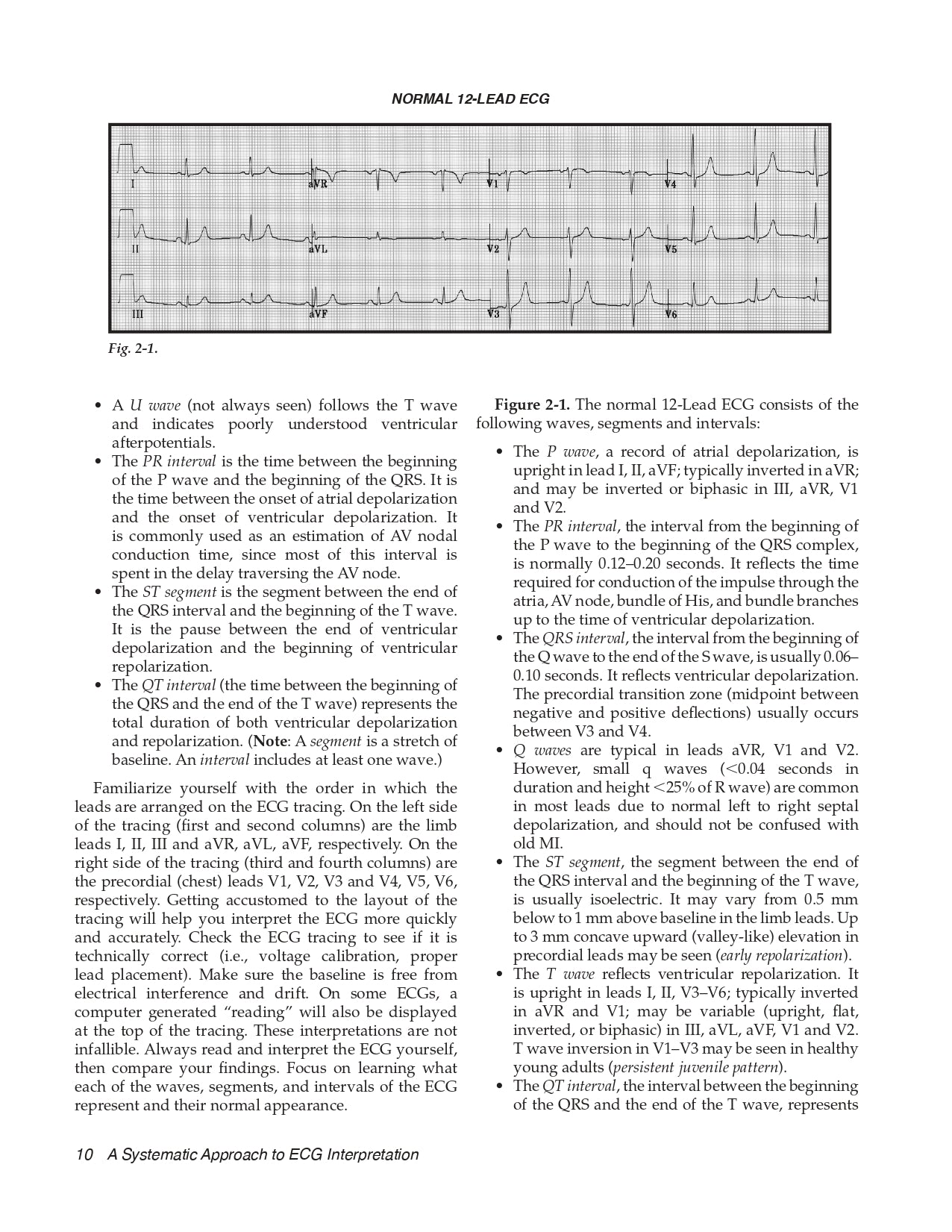
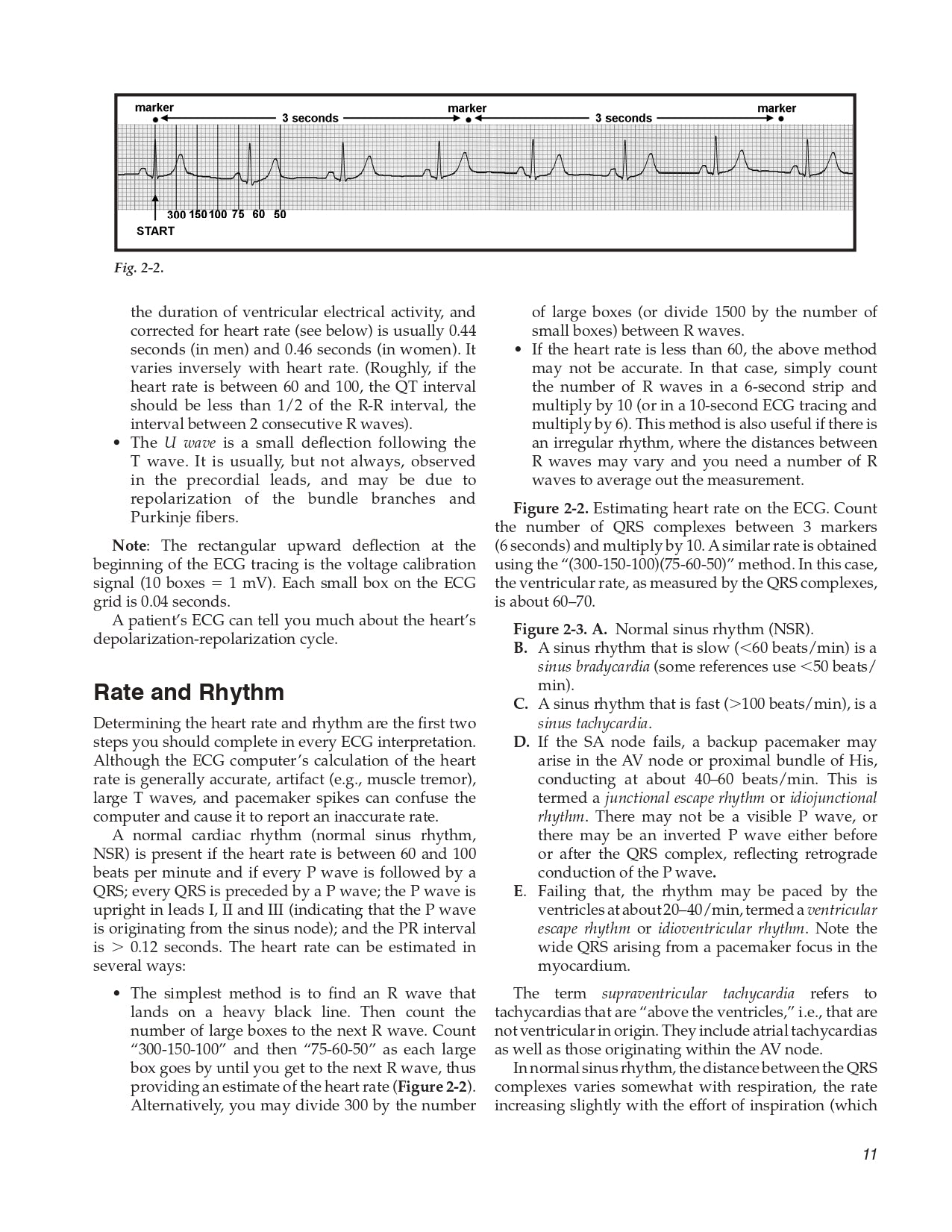
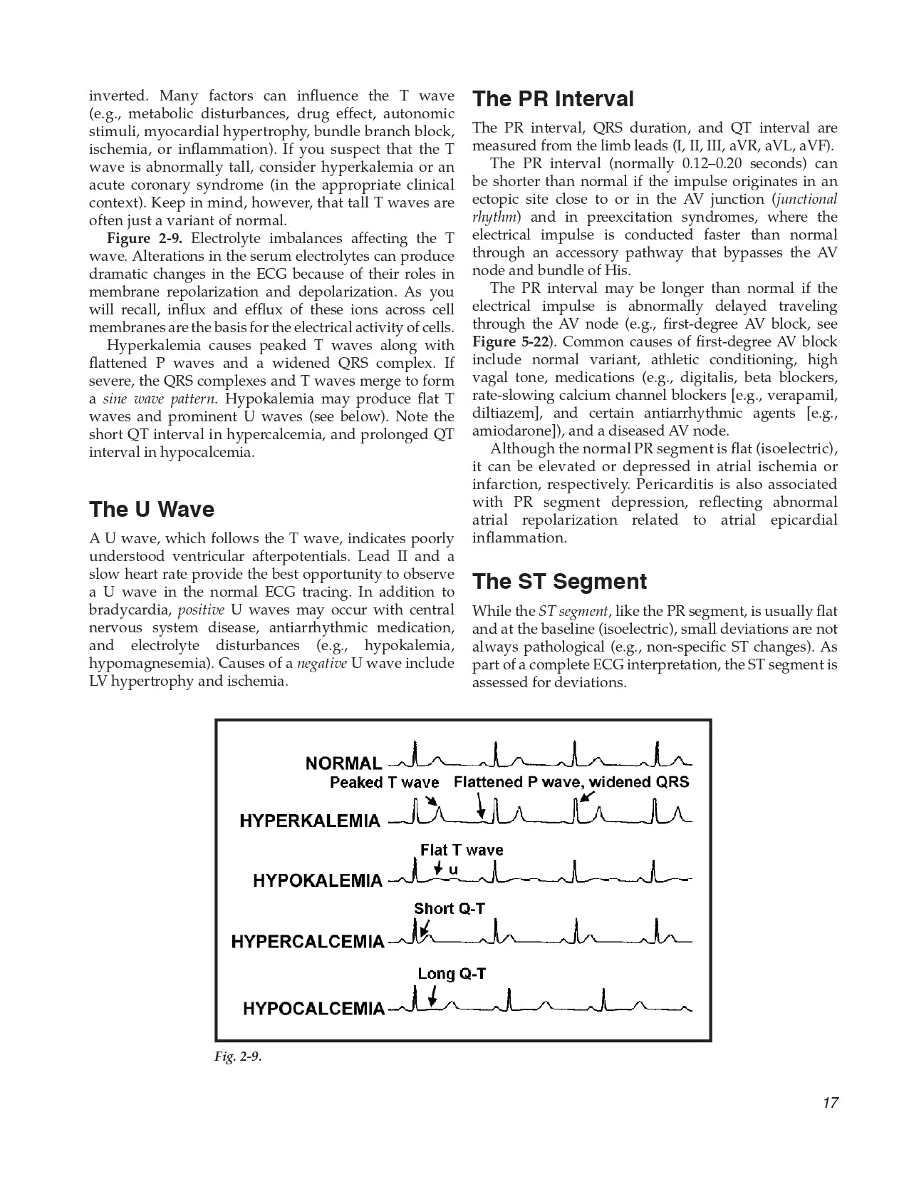
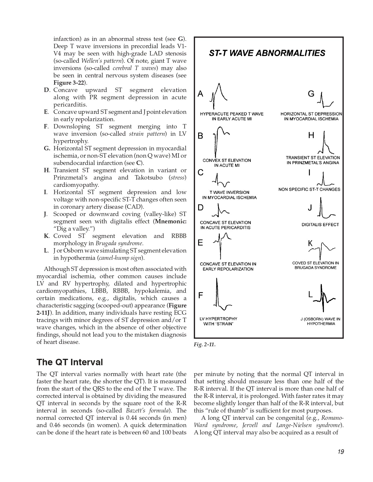
The standard 12-lead ECG • Rate and rhythm • The P wave • The QRS complex • The T wave • The U wave • The PR interval • The ST segment • The QT interval • QRS axis
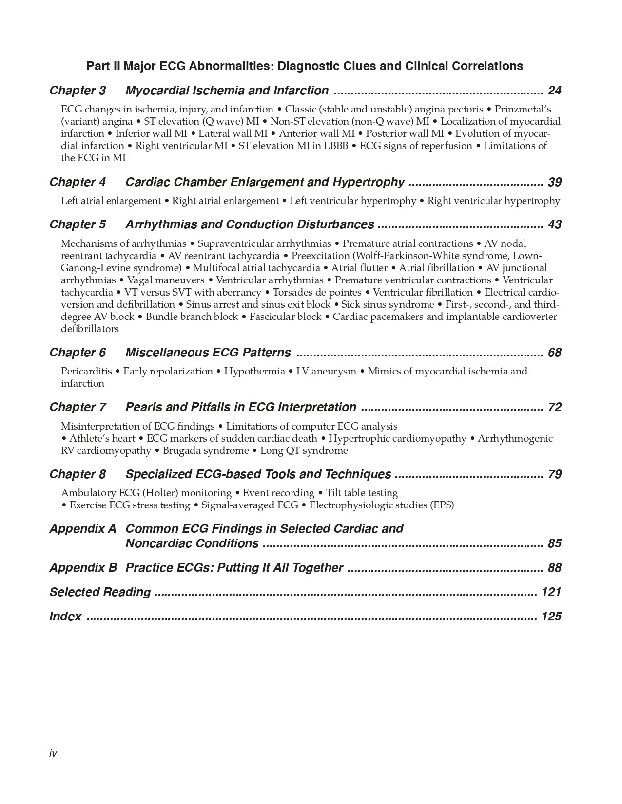
Part II Major ECG Abnormalities: Diagnostic Clues and Clinical Correlations
Chapter 3 Myocardial Ischemia and Infarction
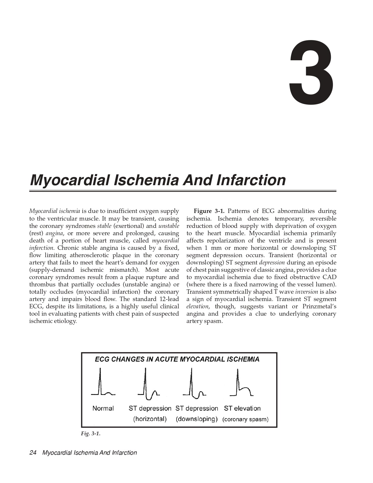
ECG changes in ischemia, injury, and infarction • Classic (stable and unstable) angina pectoris • Prinzmetal’s (variant) angina • ST elevation (Q wave) MI • Non-ST elevation (non-Q wave) MI • Localization of myocardial infarction • Inferior wall MI • Lateral wall MI • Anterior wall MI • Posterior wall MI • Evolution of myocardial infarction • Right ventricular MI • ST elevation MI in LBBB • ECG signs of reperfusion • Limitations of the ECG in MI
Chapter 4 Cardiac Chamber Enlargement and Hypertrophy
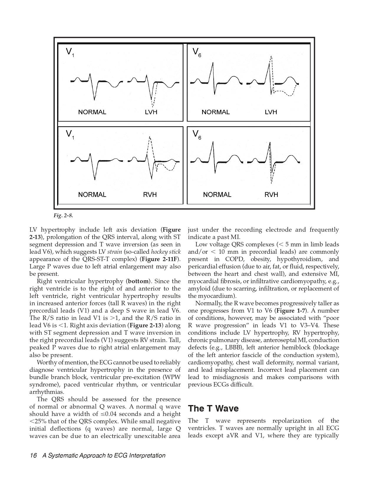
Left atrial enlargement • Right atrial enlargement • Left ventricular hypertrophy • Right ventricular hypertrophy
Chapter 5 Arrhythmias and Conduction Disturbances
Mechanisms of arrhythmias • Supraventricular arrhythmias • Premature atrial contractions • AV nodal reentrant tachycardia • AV reentrant tachycardia • Preexcitation (Wolff-Parkinson-White syndrome, Lown-Ganong-Levine syndrome) • Multifocal atrial tachycardia • Atrial flutter • Atrial fibrillation • AV junctional arrhythmias • Vagal maneuvers • Ventricular arrhythmias • Premature ventricular contractions • Ventricular tachycardia • VT versus SVT with aberrancy • Torsades de pointes • Ventricular fibrillation • Electrical cardioversion and defibrillation • Sinus arrest and sinus exit block • Sick sinus syndrome • First-, second-, and third- degree AV block • Bundle branch block • Fascicular block • Cardiac pacemakers and implantable cardioverter defibrillators
Chapter 6 Miscellaneous ECG Patterns
Pericarditis • Early repolarization • Hypothermia • LV aneurysm • Mimics of myocardial ischemia and infarction
Chapter 7 Pearls and Pitfalls in ECG Interpretation
Misinterpretation of ECG findings • Limitations of computer ECG analysis • Athlete’s heart • ECG markers of sudden cardiac death • Hypertrophic cardiomyopathy • Arrhythmogenic RV cardiomyopathy • Brugada syndrome • Long QT syndrome
Chapter 8 Specialized ECG-based Tools and Techniques
Ambulatory ECG (Holter) monitoring • Event recording • Tilt table testing • Exercise ECG stress testing • Signal-averaged ECG • Electrophysiologic studies (EPS)
Appendix A Common ECG Findings in Selected Cardiac and Noncardiac Conditions
Appendix B Practice ECGs: Putting It All Together
Selected Reading
Index


